
QT Prolongation Risk Estimator
Assess the relative risk of QT interval prolongation by selecting an antibiotic and identifying patient-specific risk factors. This tool helps guide decisions on whether baseline or follow-up ECG monitoring is necessary.
Recommendation
You prescribe a course of antibiotics for a stubborn infection, feeling confident in your choice. But have you checked the patient's heart rhythm lately? It might sound like an overreaction, but combining common antibiotics like fluoroquinolones or macrolides with certain patient profiles can trigger QT interval prolongation, a subtle electrical delay that occasionally spirals into fatal arrhythmias. This isn't just theoretical risk; it's a real clinical hazard that demands more than a cursory glance at the drug label. If you're managing patients on these agents, especially those with underlying health issues, understanding how to monitor effectively is non-negotiable.
Why Your Heart's Electrical Clock Matters
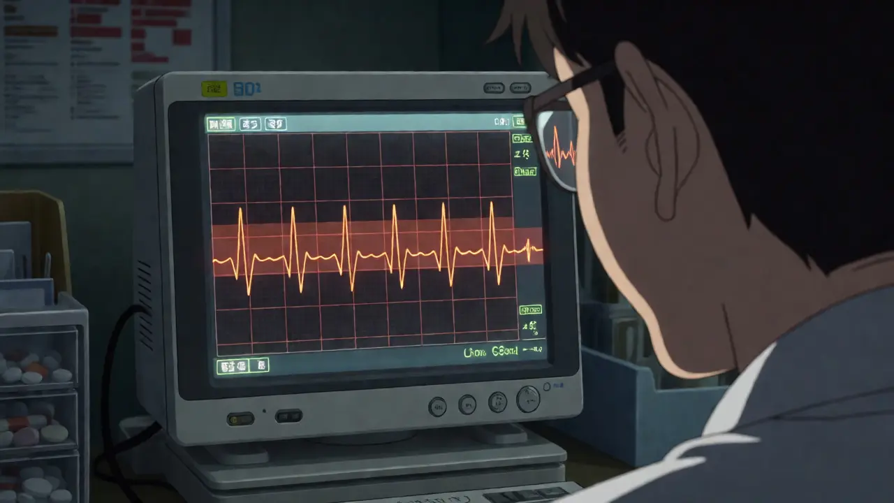
The QT interval represents the time it takes for your heart's ventricles to recharge after each beat. Think of it as resetting a clock before the next tick. When drugs interfere with this reset process-specifically by blocking potassium channels known as hERG-the clock slows down. This delay is called QT prolongation. Most of the time, a slightly longer QT interval doesn't cause immediate trouble. However, if it stretches too far, it creates a vulnerable window where chaotic electrical signals can start firing randomly. The most dangerous outcome here is Torsades de Pointes (TdP), a specific type of ventricular tachycardia that can lead to sudden cardiac death if not caught quickly.
Both fluoroquinolones (like ciprofloxacin and levofloxacin) and macrolides (such as azithromycin and erythromycin) are well-known culprits. They don't usually cause problems in healthy young people taking short courses. The danger spikes when you stack multiple risk factors together. Imagine an elderly woman with low potassium levels, already taking an antidepressant that affects heart rhythm, now starting a week of levofloxacin for a urinary tract infection. That combination creates a perfect storm for electrical instability.
Not All Antibiotics Are Created Equal
If you think all fluoroquinolones carry the same cardiac risk, you're missing key distinctions. There's a clear hierarchy of danger among these drugs. Historically, sparfloxacin was so risky it got pulled from the market. Today, we know that moxifloxacin carries a higher risk of QT prolongation compared to levofloxacin, which in turn is riskier than ciprofloxacin. Ciprofloxacin is often considered to have minimal risk for QTc prolongation in standard doses, making it a safer bet for patients with borderline cardiac health.
The same logic applies to macrolides. Erythromycin acts similarly to class III antiarrhythmic drugs and poses a significant threat. Clarithromycin sits in the middle, while azithromycin generally has the lowest impact on the QT interval among the common macrolides. Choosing azithromycin over erythromycin for a patient with existing heart conditions isn't just a preference; it's a strategic move to lower arrhythmia risk.
| Antibiotic Class | Drug Example | Relative QT Risk | Key Considerations |
|---|---|---|---|
| Fluoroquinolones | Moxifloxacin | High | Often used for respiratory infections; highest risk in this class. |
| Levofloxacin | Moderate | Commonly prescribed; requires caution in elderly patients. | |
| Ciprofloxacin | Low/Minimal | Generally safer for QT interval; preferred in high-risk groups. | |
| Macrolides | Erythromycin | High | Strong hERG channel inhibition; similar to amiodarone effects. |
| Clarithromycin | Moderate | Metabolized by liver enzymes that may interact with other drugs. | |
| Azithromycin | Low | Long half-life but lower direct electrophysiological impact. |
Who Is Actually at Risk?
It's easy to assume only patients with diagnosed heart disease need monitoring. In reality, the list of risk factors is broader and sneakier. Age is a major one. Women over 65 are particularly vulnerable, partly because they naturally have longer baseline QT intervals than men. Add in electrolyte imbalances-low potassium or magnesium-and the risk multiplies. These minerals are critical for stabilizing the heart's electrical activity. If a patient is vomiting or on diuretics, their electrolytes might be dangerously low without obvious symptoms.
Then there's polypharmacy. Many patients take other medications that also prolong QT, such as certain antipsychotics, antidepressants, or antiemetics. When you add an antibiotic to that mix, you're compounding the effect. Critically ill patients are the most susceptible group. A 2021 study highlighted that ICU patients often have multiple concurrent risk factors, making them highly prone to developing prolonged QTc intervals even with standard antibiotic doses. If you're treating someone in the hospital who is septic, hypothermic, or bradycardic, their threshold for arrhythmia is much lower.

How to Measure QT Correctly
You can't just look at a raw QT measurement on an ECG printout and make a decision. Heart rate affects QT duration-faster rates shorten it, slower rates lengthen it. To compare apples to apples, clinicians use corrected QT formulas. The two main ones are Bazett's and Fridericia's.
Bazett's formula (QTc = QT / √RR) is widely used but flawed. It tends to overcorrect at high heart rates and undercorrect at low ones, potentially giving false alarms or missing real risks. The Fridericia formula (QTc = QT / ∛RR) is increasingly preferred because it provides better accuracy across different heart rates and has been shown to predict mortality outcomes more reliably. If your ECG machine defaults to Bazett, check if you can switch settings or manually calculate using Fridericia for critical decisions.
Be aware of technical pitfalls too. Bundle branch blocks or paced rhythms can artificially inflate QT measurements. If a patient has a left bundle branch block, their QRS complex is wide, which distorts the QT reading. In these cases, interpreting the QT interval requires specialized knowledge or alternative assessment methods. Don't panic if the number looks high; first, rule out conduction abnormalities.
A Practical Monitoring Protocol
So, when exactly should you order an ECG? You don't need to scan every patient's heart before every dose. Instead, adopt a risk-stratified approach. For patients with no history of QT prolongation and no additional risk factors, routine ECG monitoring might not be necessary unless new risk factors emerge during treatment.
However, for those with identified risks, follow a structured timeline:
- Baseline ECG: Always obtain this before starting therapy in high-risk patients. This gives you a reference point to measure changes against.
- Early Check: Perform a second ECG 7-15 days after initiating fluoroquinolones or one month after starting long-term macrolide therapy. This captures the period where drug accumulation or metabolic interactions are most likely to manifest.
- Ongoing Surveillance: For patients on chronic macrolide therapy, monthly checks for the first three months are recommended by guidelines like those from the British Thoracic Society. Afterward, periodic monitoring suffices if stability is confirmed.
Timing matters. Drug concentrations peak at different times. For some antibiotics, checking the ECG roughly two hours after administration captures the maximum effect. Continuous telemetry in hospital settings offers the best data, allowing you to see fluctuations throughout the day rather than a single snapshot.
When to Stop and What to Do Next
Detecting prolongation is step one; acting on it is step two. A general rule of thumb is that a QTc interval exceeding 500 ms is clinically significant and warrants intervention. An increase of more than 60 ms from the patient's baseline is also a red flag, even if the absolute number stays below 500 ms.
If you spot these thresholds, stop the offending antibiotic immediately. Switching to an agent with lower cardiac risk, like ciprofloxacin instead of moxifloxacin, or azithromycin instead of erythromycin, is often the best move. Simultaneously, correct reversible causes. Repleting potassium to above 4.0 mmol/L and magnesium to above 2.0 mg/dL can stabilize the myocardium rapidly. Never ignore electrolytes-they are the cheapest and fastest way to reduce arrhythmia risk.
Documentation is crucial. Record the specific QTc value, the formula used, and the clinical context. This protects you and helps future providers understand the patient's sensitivity. If Torsades de Pointes occurs, treat it as a medical emergency with magnesium sulfate and pacing if needed.
Frequently Asked Questions
Is QT prolongation always symptomatic?
No, QT prolongation itself is typically asymptomatic. Patients usually don't feel anything until a serious arrhythmia like Torsades de Pointes develops, which can present as dizziness, palpitations, syncope (fainting), or sudden cardiac arrest. This silent nature makes routine monitoring essential for at-risk populations.
Which antibiotic is safest for patients with existing heart conditions?
Among fluoroquinolones, ciprofloxacin generally carries the lowest risk for QT prolongation. Among macrolides, azithromycin is typically preferred over erythromycin or clarithromycin due to its lower affinity for hERG potassium channels. However, individual patient factors and concurrent medications must always be evaluated.
Do I need an ECG for every patient prescribed these antibiotics?
Not necessarily. Guidelines suggest risk-stratified monitoring. Healthy patients with no history of cardiac issues and no other QT-prolonging drugs may not require routine ECGs. However, baseline ECGs are strongly recommended for elderly patients, women, those with electrolyte disturbances, or anyone taking multiple QT-affecting medications.
What is the difference between Bazett and Fridericia formulas?
Bazett's formula adjusts QT based on heart rate but can overestimate risk at high heart rates and underestimate it at low rates. Fridericia's formula uses a cubic root correction, providing more accurate results across varying heart rates and better predicting adverse outcomes. Fridericia is increasingly favored in clinical practice for precision.
Can correcting electrolytes reverse QT prolongation?
Yes, correcting low potassium and magnesium levels can significantly shorten the QT interval and reduce the risk of arrhythmias. Targeting potassium levels above 4.0 mmol/L and magnesium above 2.0 mg/dL is a standard intervention when QT prolongation is detected during antibiotic therapy.
Next Steps for Clinicians
Review your current prescribing habits. Are you defaulting to moxifloxacin for pneumonia without checking the patient's medication list? Are you ignoring baseline ECGs in elderly UTI patients? Small changes in protocol can prevent rare but catastrophic events. Start by identifying your high-risk cohort-patients over 65, those on polypharmacy, and those with electrolyte vulnerabilities. Implement a simple checklist for ECG timing. And remember, when in doubt, choose the antibiotic with the lower cardiac profile and verify with a quick ECG. Your vigilance protects your patients from silent electrical threats.








Write a comment
Your email address will be restricted to us